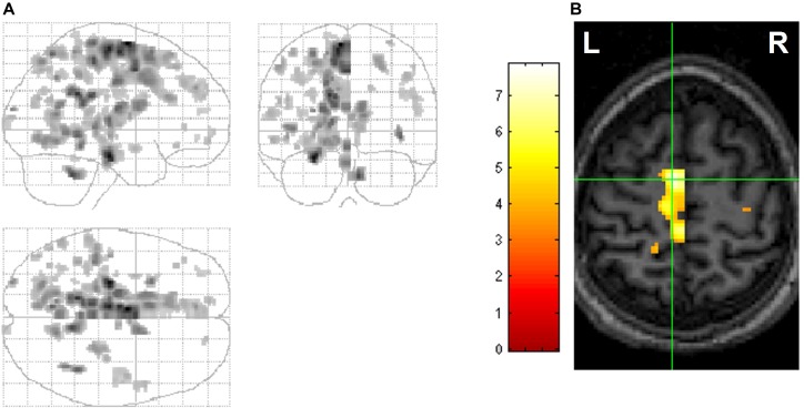
FIGURE 6.
Hemispheric differences in REM-locked activation. Voxels with greater REM-locked activity compared to its homolog in the other hemisphere are shown. All images are thresholded at uncorrected P < 0.001 (T = 3.5) and additionally at a spatial extent of >5 contiguous voxels. (A) Orthogonally oriented statistical parametric maps (maximum intensity projections). REM-locked activation was greater in the posterior left hemisphere. (B) Cross at the greatest hemispheric difference, supplementary eye field (SEF) (left greater than right). The left supplementary eye field has a clearly predominant role in sequencing saccade programming during visual scanning in wakefulness. Adapted from Hong et al. (2009) with the permission from Hong.

