. 2018 Jul 31;7(6):1144–1152. doi: 10.1039/c8tx00093j

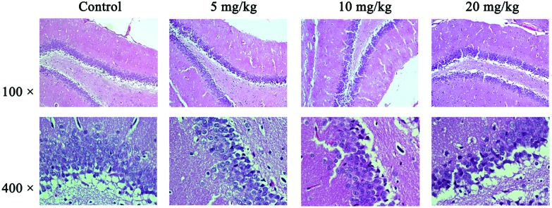
Fig. 2. Pathological results in the hippocampal tissues. The results were detected by HE staining and observed under an optical microscope. The changes in tissue histopathology were observed under a light microscope (Nikon Eclipse 80i, Tokyo, Japan) and photos were taken (×100 and ×400, respectively, for each group). Shown are the representative images for the control group (a), 5 mg kg–1 group (b), 10 mg kg–1 group (c), and 20 mg kg–1 group (d), respectively. Neurons presented pyknosis and the blurred boundary between the nucleus and cytoplasm for partial nucleus neurons in the hippocampal CAI area can be seen in the 10 mg kg–1 group. Neurons with nuclear condensation and slight edema in the hippocampus can be seen in the 20 mg kg–1 group.

Fig. 2