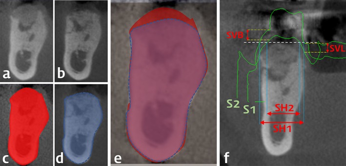
Figure 3.

Representative radiographs and overlapped 3D‐scanned study casts (the same case with Figure 1) and linear measurements. Cross‐sectional view from computed tomography taken at the time of surgery (A and C) and 4 months after the surgery (B and D). In the automatically superimposed images from the two time‐points (E), vertical and horizontal changes of the alveolar bone dimension were measured. Surface information from the 3D‐scanned study casts (S1 at the time of surgery; S2 at 4 months after the surgery) were superimposed with the computed tomography at 4 months after the surgery (F). In this view, horizontal soft tissue dimension was measured at the most‐crestal level of the regenerated alveolar bone at two time‐points (SH1 and SH2), and horizontal reduction was calculated by their subtraction. Vertical change of soft tissue dimension was measured at both buccal and lingual gingival margins around the extracted tooth (SVB and SVL)
