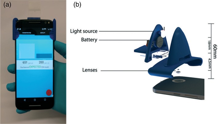
Figure 4.

(a) Picture of a smartphone‐based cytometer with the microfluidic chip mounted on it. It is worth noting the compactness of the attached plastic system, (b) schematic view of the external elements needed to perform the bright field microscopy: An external light source and its battery for sample illumination and the optical lens to create the microscope together with the lens already present in the mobile phone (reproduced from Ref. 95 with permission from The Royal Society of Chemistry). [Color figure can be viewed at http://wieyonlinelibrary.com]
