
Figure 3.
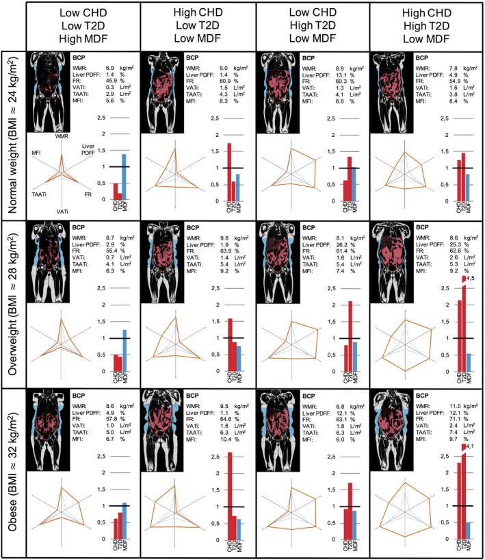
Body composition profiling of females from the UK Biobank imaging cohort. Each subject, approximately age 65, is presented with a coronal slice from the MRI scan with VAT (red) and ASAT (blue) segmentations, the BCP values with corresponding six‐axes plots, and bar plots showing sex‐and‐age normalized predicted probabilities. BCP, body composition profile; CHD, coronary heart disease; FR, fat ratio; MDF, metabolic disease free; MFI, muscle fat infiltration; ASAT, abdominal subcutaneous adipose tissue; PDFF, proton density fat fraction; T2D, type 2 diabetes; TAATi, total abdominal adipose tissue index; VATi, visceral adipose tissue index; WMR, weight‐to‐muscle ratio.