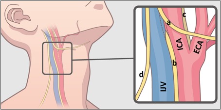
Figure 2.

Scheme of the anatomy of the human neck. Black frame represents the approximate area dissected in this study. Blue structure is internal jugular vein (IJV), red is carotid artery (ICA: internal carotid artery, ECA: external carotid artery). Yellow lines represent the nerves. (a: hypoglossal nerve, b: vagus nerve, c: glossopharyngeal nerve, d: spinal accessory nerve) [Color figure can be viewed at http://wileyonlinelibrary.com]
