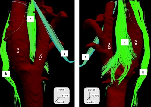
Figure 4.

The diffusion tensor tractography fusion with artery structure obtained from CT. Right side neck. Sample number 2. a, The hypoglossal nerve; b, the vagus nerve; and c, the glossopharyngeal nerve. ECA, external carotid artery; ICA, internal carotid artery [Color figure can be viewed at http://wileyonlinelibrary.com]
