Figure 5.

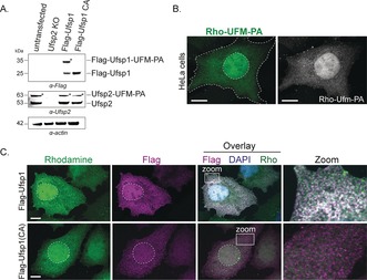
Reactivity of Hela cells electroporated with Rho‐UFM1‐PA in the absence or presence of ectopically overexpressed murine Flag‐Ufsp1. A) Immunoblots of Flag‐Ufsp1 and Flag‐Ufsp1(C53A) and endogenous Ufsp2 in untransfected cells following electroporation. B) Confocal images of untransfected HeLa cells or C) in the presence of Flag‐Ufsp1 or Flag‐Ufsp1 (C53A) after probe electroporation. Cell boundaries and nuclei are indicated by a dashed line, and insets correspond to zoom‐ins. Scale bars=10 μm. Quantification of colocalization (Mander's overlap coefficient) is shown in Figure S11.
