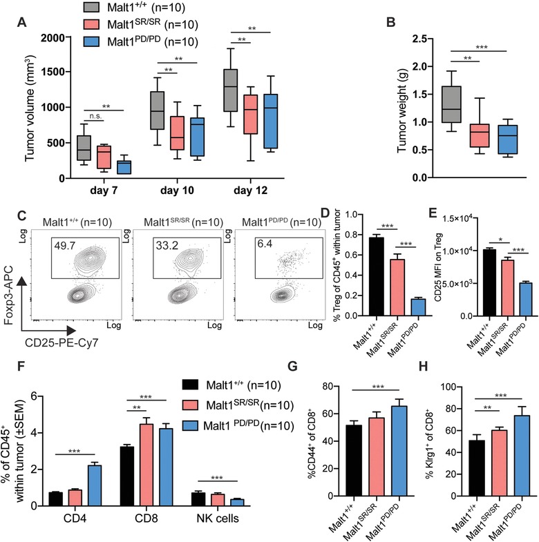
Figure 4.

Malt1‐deficiency reduces MC38 tumor growth, by improving anti‐tumor immunity due to Treg impairment (A) Tumor volume was determined by blinded repeated caliper measurements at indicated time points in animals of the different cohorts. (B) Tumor weight at end stage. (C) Frequency of CD4+ Foxp3+ regulatory T cells in the tumor microenvironment was measured by flow cytometry (Malt1+/+ n = 10, Malt1SR/SR n = 10, Malt1PD/PD n = 10). (D) Frequency of total tumor infiltrating Treg as percentage of infiltrating CD45+ lymphocytes (Malt1+/+ n = 10, Malt1SR/SR n = 10, Malt1PD/PD n = 10). (E) CD25 MFI was measured by flow cytometry on tumor infiltrating Treg(Malt1+/+ n = 10, Malt1SR/SR n = 10, Malt1PD/PD n = 10). (F) Frequency of total tumor infiltrating CD4+ Foxp3− T cells, CD8+ T cells and Nkp46+ NK cells (Malt1+/+ n = 10, Malt1SR/SR n = 10, Malt1PD/PD n = 10). (G) Expression of the activation marker CD44 and (H) Klrg1 on CD8+ T cells within the tumor microenvironment. The data are shown as mean +SD (Malt1+/+ n = 10, Malt1SR/SR n = 10, Malt1PD/PD n = 10) and are representative of three independent experiments.
