Figure 1.

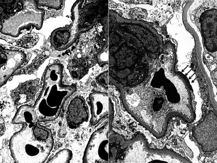
Electron microscopy (4000‐fold) of the allograft on day 19 after transplantation. Complete flattening of podocyte foot processes (black arrows). Minimal change lesion of incipient FSGS

Electron microscopy (4000‐fold) of the allograft on day 19 after transplantation. Complete flattening of podocyte foot processes (black arrows). Minimal change lesion of incipient FSGS