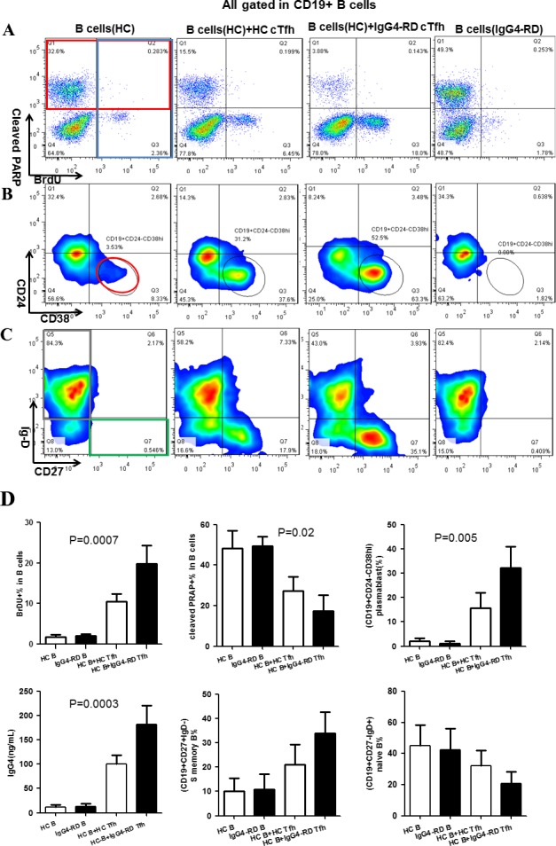
Figure 4.

Impact of circulating Tfh (cTfh) cells on B cell proliferation, apoptosis, and differentiation. Magnetic bead–purified B cells from healthy controls were cultured alone or cocultured with CD4+CXCR5+ T cells in vitro. The 4 groups were healthy control B cells alone, healthy control B cells cocultured with healthy control cTfh cells, healthy control B cells cocultured with cTfh cells from patients with IgG4‐RD, and B cells from patients with IgG4‐RD alone. Representative data from 4 independent experiments are shown. A, Detection of bromodeoxyuridine (BrdU) (blue box) and cleaved poly(ADP‐ribose) polymerase (PARP) (red box) by flow cytometry in B cells on day 3 after stimulation. B, Detection of CD19+CD24−CD38high plasmablasts (red circle) by flow cytometry on day 7 after stimulation. C, Detection of naive B cells (CD19+CD27−IgD+) (gray box) and switched (S) memory B cells (CD19+CD27+IgD−) (green box) by flow cytometry on day 7 after stimulation. D, Percentages of BrdU+ B cells, cleaved PARP+ B cells, plasmablasts/plasma cells, switched memory B cells, and naive B cells and concentrations of IgG4 in the cultured cell suspensions from the different groups. Results are the mean ± SD. See Figure 1 for other definitions.
