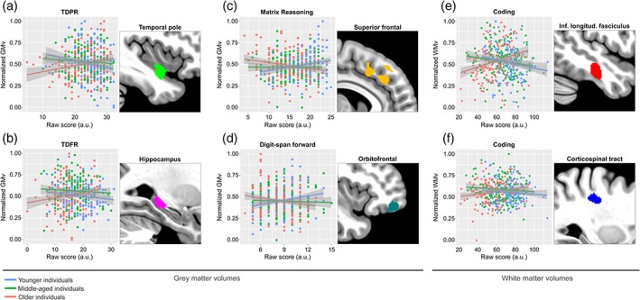
Figure 5.

Aging significantly modulated the relationships between cerebral volumes and cognitive performance. Gray matter volumes are plotted against measures of delayed paired recall (a), delayed free recall (b), nonverbal reasoning (c), and working memory (d), in the left temporal pole, left hippocampus, right superior frontal, and right orbitofrontal cortex, respectively. White matter volumes are plotted against measures of cognitive processing speed in the right inferior longitudinal fasciculus (e) and the right dorsal aspect of the corticospinal tract (f), respectively. For visualization purposes the sample was broken down into three different age categories, using tercile grouping. GMv as well as WMv values are adjusted for age, sex, years of education, and total intracranial volume (TIV). Shaded areas in the scatterplots indicate 90% confidence intervals [Color figure can be viewed at http://wileyonlinelibrary.com]
