Figure 1.

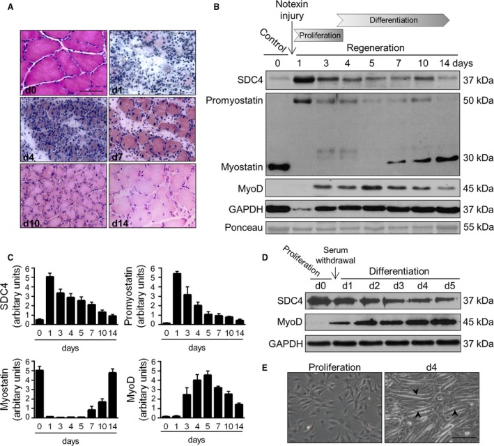
Expression of SDC4 and myostatin during skeletal muscle regeneration. (A) Representative haematoxylin and eosin‐stained sections of control and regenerating soleus muscle of the rat on different days after notexin injection. Bar: 50 μm. (B) Aliquots of extracts containing equivalent amounts of protein obtained from m. soleus on different days after notexin induced injury were subjected to SDS/PAGE, and immunoblotted with anti‐SDC4, anti‐myostatin (AB3239‐I), anti‐MyoD and anti‐GAPDH antibodies. Representative immunoblots are shown. GAPDH level is decreased after the injury in the necrotic muscle. Representative Ponceau staining of the membrane is presented. (C) Quantification of results, data are reported as means ± SEM (n = 4 independent experiments at each time point). (D) Expression of SDC4 during the differentiation of C2C12 myoblasts (0–5 days). GAPDH shows the equal loading of the samples. (E) Representative images of proliferating and differentiating (day 4) C2C12 cells. Arrowheads show the formation of myotubes. Bar: 200 μm.
