
Figure 1.
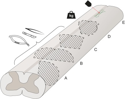
Commonly used rodent DC lesion models. A schematic diagram of the rat spinal cord and common DC lesion paradigms. Injured areas are depicted with striped lines, together with instruments commonly used to perform the lesion. (A–C) Transection injuries of the spinal cord, illustrating in (A) lateral hemisection of the spinal cord, (B) dorsal hemisection of the spinal cord, and (C) bilateral transection of the DC (microscissors and scalpel depicted, other instruments are also used as summarized in Table 1 and Supporting Information, Table 2). Besides transection, the DC lesion can be implemented using forceps creating a crush injury. (D) Contusion or compression injury by dropping or placing a weight on the spinal cord in a controlled manner. (E) Severing individual superficial DC axons using a laser. [Color figure can be viewed at http://wileyonlinelibrary.com]