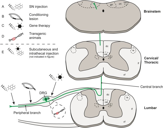
Figure 3.

The anatomy of the dorsal root ganglia, the spinal cord and targets for neuron‐intrinsic experimental intervention strategies. Axons of DRG neurons bifurcate into two branches, one going into the periphery and the other going into the spinal cord. These axons relay information including heat, pain, and position from the body and project via the DC to the brainstem directly (the gracile nucleus, cuneate nucleus and the external cuneate nucleus) or indirectly via spinal neurons (not shown). Not shown: collaterals also innervate spinal cord grey matter in the segments around where they enter, and some collaterals descend caudally. Axon collaterals also innervate Clarke's nucleus in the thoracic cord. The arrows indicate targets for neuron‐intrinsic intervention to promote axonal growth and plasticity, with (A) introduction of pharmacological agents by injection into the SN, (B) CL of the SN, (C) viral vector delivery by injection into the DRG, (D) using transgenic animals, and (E) subcutaneous or intrathecal injection to deliver pharmacological agents (not illustrated). Key: gn = gracile nucleus, cn = cuneate nucleus, ecn = external cuneate nucleus, dc = dorsal column, cst = corticospinal tract, d = dorsal nucleus (Clarke's nucleus), rs = rubrospinal tract, lf = lateral funiculus, vf = ventral funiculus. [Color figure can be viewed at http://wileyonlinelibrary.com]
