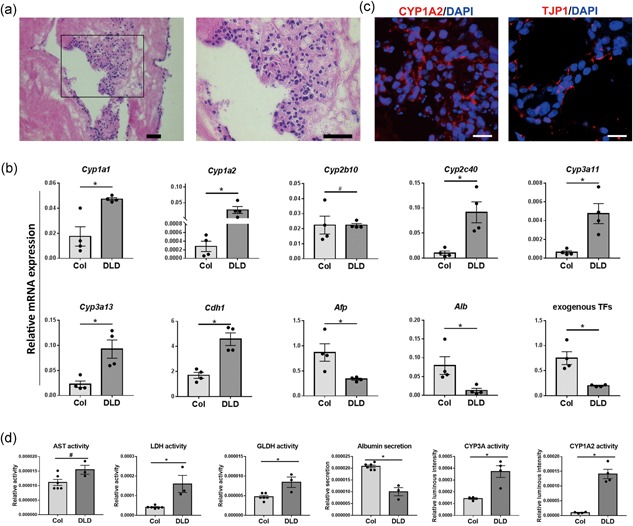
Figure 5.

Repopulation of decellularized liver disks by iHeps. (a) Hematoxylin and eosin (H&E) staining showing the repopulation of decellularized liver disks by iHeps after 1 week. Scale bar = 50 μm. (b) Immunofluorescence analysis for iHeps cultured on decellularized liver disks (DLD). Scale bar = 20 μm. (c) Gene expression analysis for iHeps cultured on collagen (type I) coated plates (Col) and DLD. (d) Relative activity of hepatocyte‐specific enzymes (AST, LDH, GLDH), CYP3A and CYP1A2 activity and albumin secretion in iHeps cultured on Col and DLD. All results were normalized with cell input with Alamar blue. Data are shown as mean ± SEM of six independent experiments for collagen 2D cultured iHeps for AST, LDH, GLDH and albumin tests, three for DLD cultured iHeps for AST, LDH, GLDH and albumin tests, four for both 2D and DLD cultured iHeps for CYP3A and CYP1A2 tests. The asterisk represents statistical significance. *p < 0.05. The hash represents nonsignificance (two‐tailed Mann–Whitney U test) [Color figure can be viewed at wileyonlinelibrary.com]
