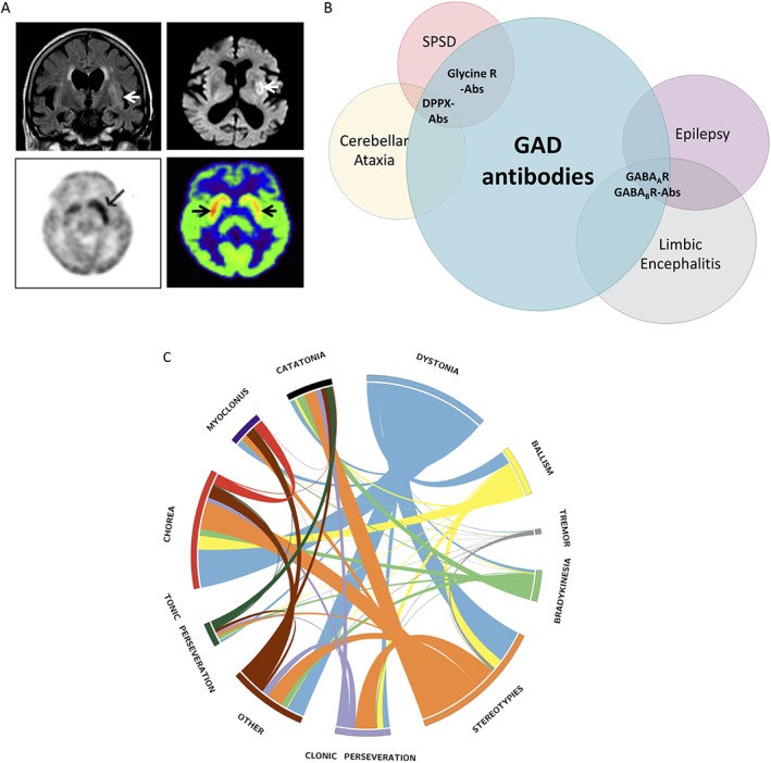
Figure 3.

Radiological spectrum of faciobrachial dystonic seizures with LGI1‐antibodies and clinical spectrum of glutamic acid decarboxylase (GAD) antibodies. (A) Multimodal radiological involvement of the basal ganglia in patients with LGI1‐antibodies and faciobrachial dystonic seizures using FLAIR and DWI‐weighted MRI (top 2 panels), PET (bottom left panel), and SPECT (bottom right panel) imaging. Arrows indicate abnormal basal ganglia regions. Reproduced with permissions.20, 49, 52 (B) Spectrum of overlapping autoimmune neurological diseases associated with GAD65 antibodies and concommittant CNS‐specific autoimmunity. Autoantibodies highlighted in bold. DPPX, dipeptidyl‐peptidase‐like protein‐6; LGI1, leucine‐rich glioma‐inactivated‐1, FLAIR, Fluid Attenuated Inversion Recovery; DWI, diffusion‐weighted imaging; SPECT, single‐photon emission computed tomography. (C) Circos diagram depicting the relative presence of phenomenological features in patients with NMDAR‐antibody encephalitis (adapted from Varley et al.29)
