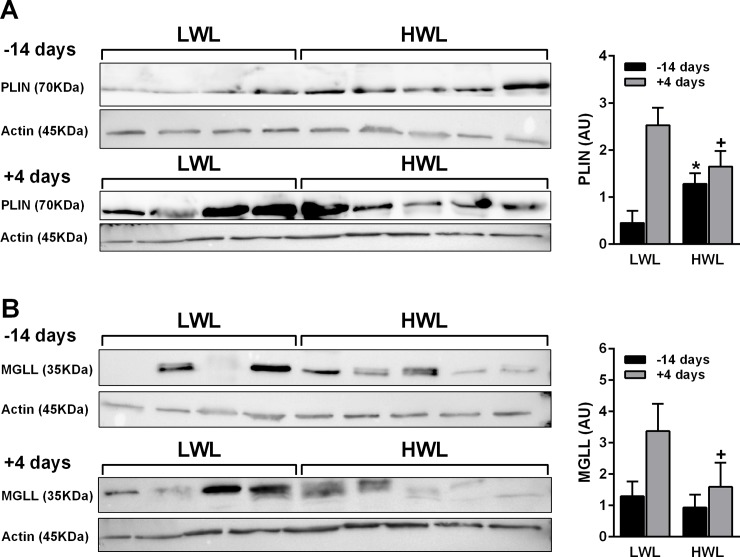
Fig 6. Protein expression of PLIN1 and MGLL in ATs collected at 14 d prepartum and 4 d PP from HWL and LWL cows.
Cows were categorized as either high-weight loss (HWL, n = 9) or low-weight loss (LWL, n = 9) based on the percentage of BW loss between weeks 1 and 5 postpartum. The protein abundances of PLIN1 (A) and MGLL (B) were assessed by Western blotting analysis and corrected by β-actin as an internal standard. Samples for the western blot were prepared (×3.5), divided, and loaded to separate gels that ran simultaneously. The MGLL, PLIN1, and β-actin ran in parallel gels due to the proximity of the bands. Data represent the mean ± SEM. *P<0.05 in HWL vs. LWL AT prepartum. +P<0.1 in HWL vs. LWL AT postpartum.

