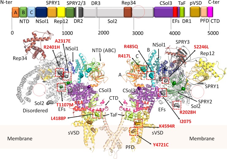
Fig 1. Domain architecture of the cardiac RyR2.
Domains are coloured according to the linear sequence scheme shown above the ribbon diagram. Structure obtained from PDB: 5GOA. [15] The ribbon diagram shows a RyR2 dimer from the side. Red circles represent large alpha solenoid regions which are unstructured in the cryo-electron micrograph (CryoEM) structure. All variants discussed in the manuscript are shown in sphere representations. Red squares highlight location of the most likely damaging variants (expanded in Fig 2). Domains are named according to nomenclature used by des Georges, et al., 2016. A (dark orange), B (green), and C (light blue) domains: form part of the N-terminal domain (NTD); NSol (dark blue): alpha-solenoid region near the NTD; SPRY1 (light orange), SPRY2 (dark green), and SPRY3 (dark grey): three domains named after splA kinase and RyRs where they were first identified; Sol2 (light grey): second alpha-solenoid region centrally located on RyR2; Rep12 (yellow) and Rep23 (brown): four repeats (~100 aa each) in two tandem arrangements, Repeats 1 and 2 located between SPRY 1 and SPRY2, and Repeats 3 and 4 located within Sol2; CSol3 (magenta): third alpha-solenoid region located near the C-terminal; EFs (red): pair of EF hand-like motifs located within CSol3 region; TaF (light green): thumb and forefingers domain; DR1/2/3 (white): evolutionary divergent regions of RyR isoforms (not shown in the ribbon diagrams); pVSD (gold): pseudo voltage-sensing domain; PFD (wheat): pore-forming domain; CTD (pink): C-terminal domain.

