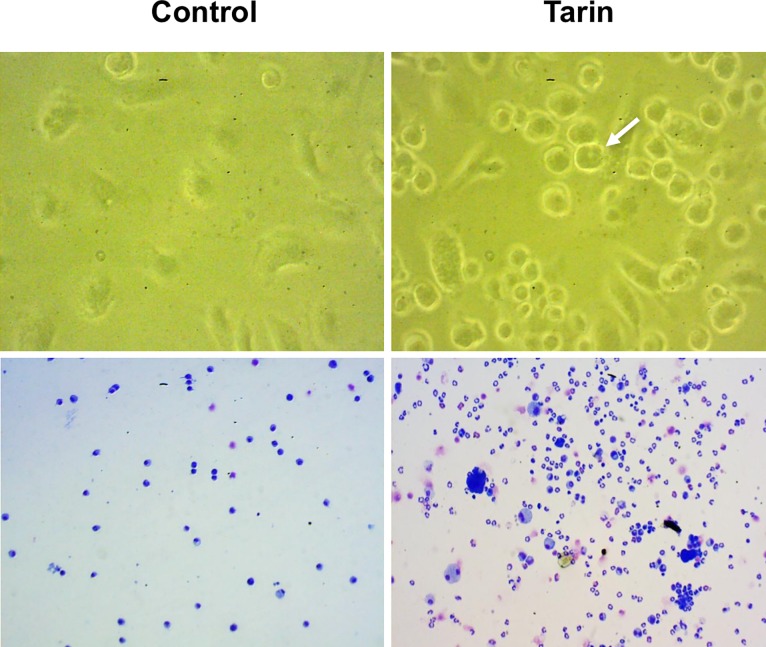
Fig 2. Morphological characteristics of bone marrow cells cultured with tarin.
BM cell cultures incubated with tarin 20 μg/mL (Tarin) and without tarin (Control) for 6 days (top panels) and 19 days (bottom panels). The white arrow indicates a typical nucleus of a myeloid cell in development. The bottom panel shows granulocytic lineage cells in distinct developmental stages in tarin-treated cells (right-hand side) or the control group (left-hand side). Photomicrographs were acquired by an inverted-phase microscopy under 400x magnification (top panel) or by visualization of cytosmears, stained by May-Grunwald Giemsa, using optical microscopy under 200x magnification (bottom panel).

