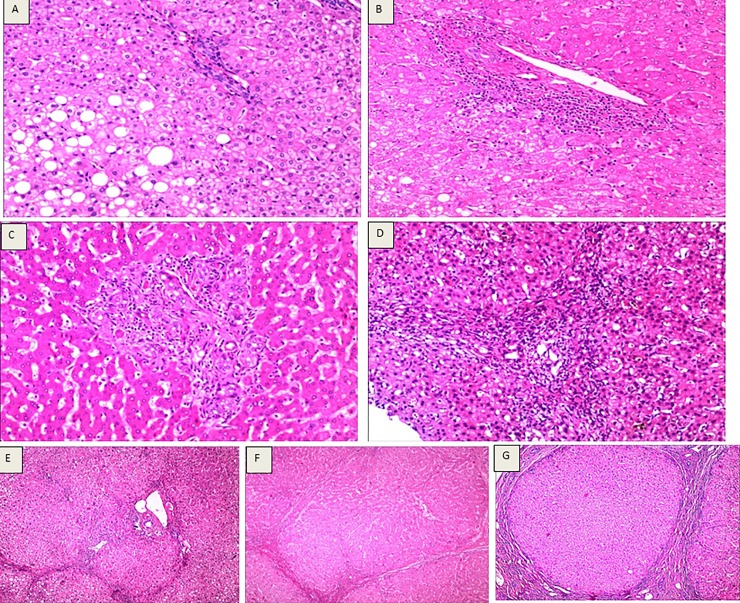
Fig 2. Assessments of liver fibrosis and cirrhosis according to the Laennec staging system.
(A) Laennec staging 0, HE staining (10×10), hepatocytic loosening, steatosis, focal necrosis, mild lymphocytic infiltration in the portal area, and no fibrosis. (B) Laennec staging 1, HE staining (4×10), loose liver cell cytoplasm, fatty degeneration, focal point necrosis, mild fiber hyperplasia and enlargement, no fiber spacing, lymphocyte infiltration within the portal area. (C) Laennec staging 2, HE staining (4×10), hepatocyte eosinophilic degeneration, eosinophilic body formation, focal necrosis, enlargement of fibrous tissue in the portal area, formation of fibrous septa, infiltration of lymphocytes, and hyperplasia of small bile ducts. (D) Laennec staging 3, HE staining (4×10), liver cell cytoplasm of osteoporosis, eosinophilic degeneration, cholestasis, focal necrosis and severe periportal piecemeal necrosis, hyperplasia of fibrous tissue expansion, formation of fibrous septa, segmentation of hepatic lobule, periportal and septa lymphocytes. (E) Laennec staging 4A, HE staining (4×10). Hepatic lobule structure disappearance, hepatocyte loosening, steatosis, focal necrosis, fibrous tissue hyperplasia, fibrous septum, formation of false lobules, infiltration of lymphocytes in the portal area and intercellular septum. (F) Laennec staging 4B, HE staining (4×10), hepatic lobule structure disappearance, liver cell vacuolization, focal necrosis and moderate periportal piecemeal necrosis, hyperplasia of fibrous tissue formation of fibrous septa, pseudolobule formation, changes in part of the fiber interval width, portal area and fibrous septum lymphocytic infiltration. (G) Laennec staging 4C, HE staining (4 x 10), hepatic lobule structure disappearance, liver cell vacuolization, focal necrosis, portal fibrosis formation of fibrous septa, fibrous septum wrapped around the liver cell cluster, formation of pseudolobules, fiber spacing width changes, portal area and fibrous septum lymphocytic infiltration).

