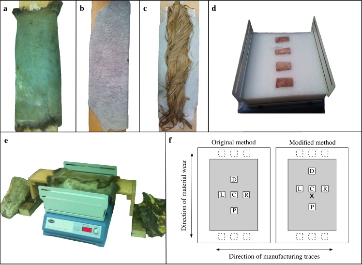
Fig 2. Setup of experiment and scanning locations.
(a-c) Material setup on external platform; (a) fresh skin; (b) processed leather; (c) dry bark; (d) bone specimens inserted into foam on a Stuart SSL2 rocking machine (bones in this image are from a test sample with placement in the foam perpendicular to the direction of all the bones in the subsequent experiment); (e) platform with material over the Stuart SSL2 in the experimental position; (f) examples of crosswise sampling using two methods for obtaining scans at locations C (center), D (distal), P (proximal), L (left), and R (right). Additional scanned locations are represented by dashed boxes at the extreme distal and proximal ends. All scanned areas are 0.8mm2 but are enlarged here for readability. Modified sampling method uses an incised “x-mark” for finding the center over all samples.

