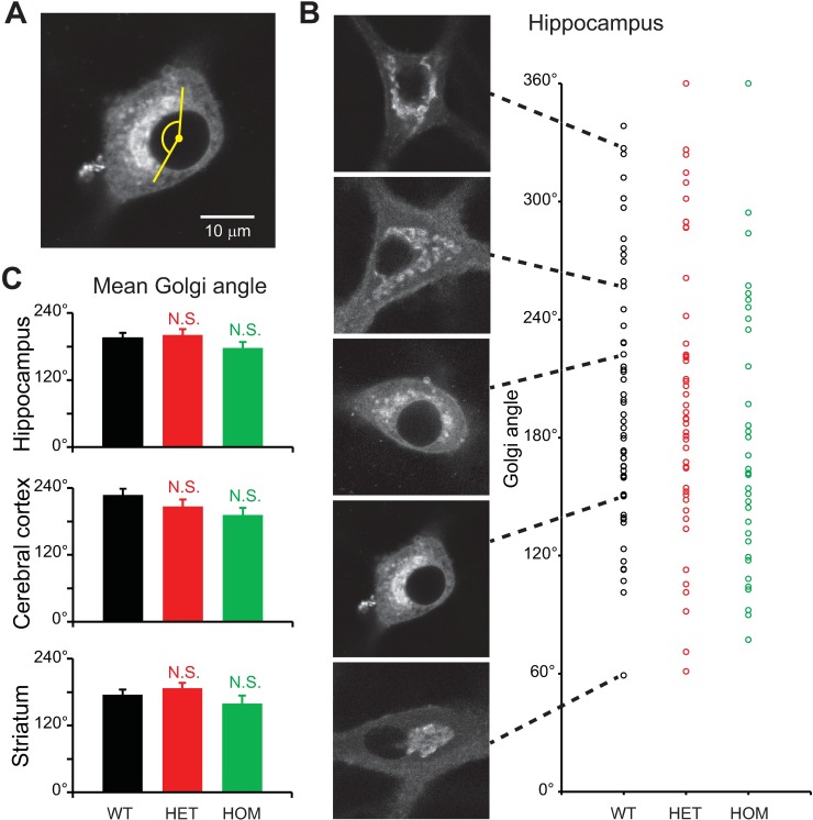
Fig 8. Degree to which the Golgi apparatus encircled nuclei.
Confocal microscopy of neurons stained with BODIPY FL C5-ceramide, carried out at 17–19 DIV. A: Representative confocal image of a WT hippocampal neuron. The extent to which the Golgi apparatus encircled the nucleus was measured as the angle subtended by the Golgi, with the center of the nucleus defined as the vertex. B: Variability of angle subtended by the Golgi in hippocampal neurons. Each circle represents a single neuron. C: Differences in values measured for mutant vs. WT neurons were not significantly different (p>0.1; t-test; n = 54, 49, 37 hippocampal neurons, 49, 40, 25 cerebral cortical neurons, and 53, 58, 29 striatal neurons for WT, HET and HOM, respectively).

