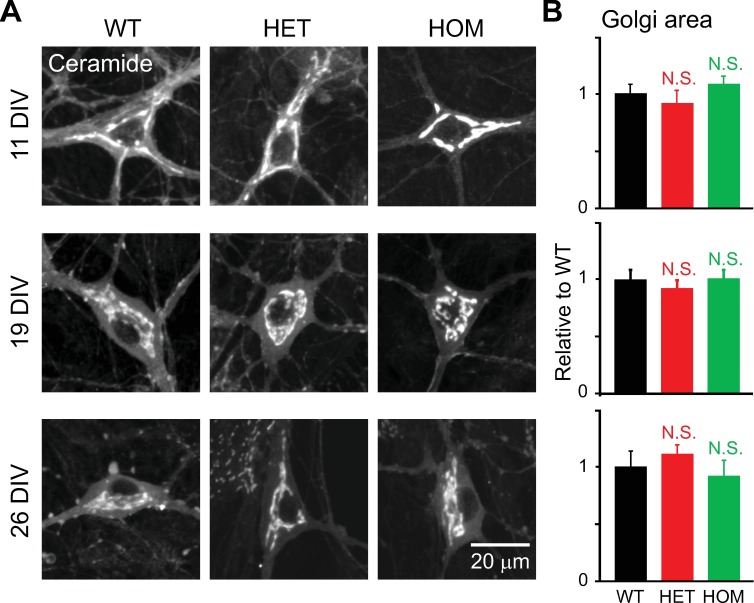
Fig 11. Structure of the Golgi apparatus during the course of culture.
Hippocampal neurons of ΔE-torsinA knock-in mice were stained with BODIPY FL C5-ceramide, and imaged by confocal microscopy at the specified DIV. A: Representative confocal images of neurons obtained from WT, HET and HOM ΔE-torsinA knock-in mice, at 11, 19 or 26 DIV. MIP images are shown. B: Differences in Golgi area between mutant and WT neurons, as analyzed by the MIP method, were not statistically significant during maturation (p>0.1; t-test; n = 26, 14, 18 11-DIV neurons, 30, 44, 32 19-DIV neurons, and 11, 17, 18 26-DIV neurons for WT, HET and HOM, respectively). All values measured were normalized to the average WT value.

