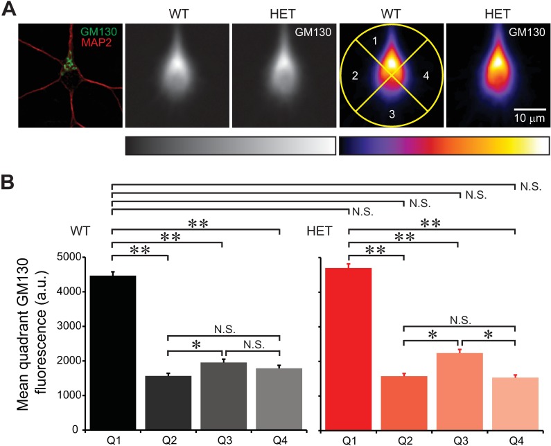
Fig 13. Degree of Golgi polarization at the base of thickest dendrite.
Hippocampal neurons were stained for GM130 at 11 DIV, centered, and oriented such that the thickest dendrite pointed toward 12 o'clock, and averaged. The polarity of the Golgi was analyzed as the distribution of GM130 intensity in the top quadrant (90° pie) relative to the other quadrants. A: The leftmost panel shows the image of a single neuron, stained by immunocytochemistry for GM130 and a dendritic marker MAP2 (single confocal plane). Two grayscale images show averaged GM130 signals in neurons from WT and HET neurons (slide scanner images). Two images on the right show the same GM130 signals shown in pseudocolor. The center of each panel corresponds to the somatic center. Four quadrants are illustrated with a yellow circle in one panel. The scale bar applies to all panels. Rectangles below the images show the corresponding color lookup tables. B: Relative distributions of the Golgi in the four quadrants. Golgi was polarized in the top quadrant (Quadrant 1, Q1) in WT and HET neurons (**: p<1.01 × 10−46; *: p<1.77 × 10⁻³). However, differences in polarization between mutant and WT neurons were not statistically significant (N.S.; p>2.61 × 10⁻² with α = 1.78 × 10⁻³ after Bonferroni correction; t-test; n = 410 and 407 neurons for WT and HET, respectively).

