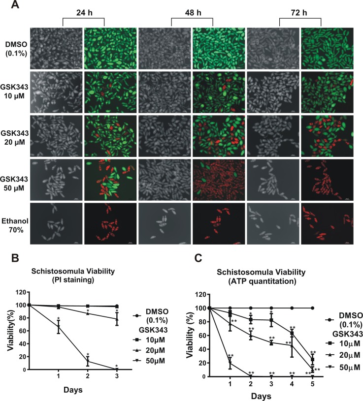
Fig 6. Quantitation of the decrease in viability of schistosomula caused by GSK343 treatment at different concentrations and incubation times.
(A) Schistosomula treated with the indicated concentrations of GSK343 or with vehicle (0.1% DMSO) for 24, 48 and 72 h were visualized by staining with propidium iodide (marker of dead cells; 572 nm emission filter microscope) and with fluorescein diacetate (marker of living cells; 492 nm emission filter microscope). The bottom row shows a positive control, namely exposure to 70% ethanol, which kills all parasites. For each time point (indicated at the top), the left panel shows a light microscopy image and the right panel shows the image of the same field with differential fluorescence detection of PI-positive dead and FDA-positive live schistosomula by superimposition of 536nm and 494nm epifluorescence spectra. (B) Quantitation of viability of treated schistosomula. Percentage of viable schistosomula (non-stained with propidium iodide) over three days of treatment. For each condition tested, about 3600 schistosomula were used, divided into four biological replicates and three time points analyzed. Mean ± SD from four replicate experiments. (C) ATP quantitation using a luminescent assay to assess schistosomula survival under GSK343 exposure. Schistosomula (100-120/well) were incubated with the indicated concentrations of GSK343 or with vehicle (0.1% DMSO) for up to 5 days. The viability was expressed as % the luminescence values relative to the control (DMSO). Mean ± SD from three replicate experiments. *p < 0.05; **p < 0.01 (two-way ANOVA).

