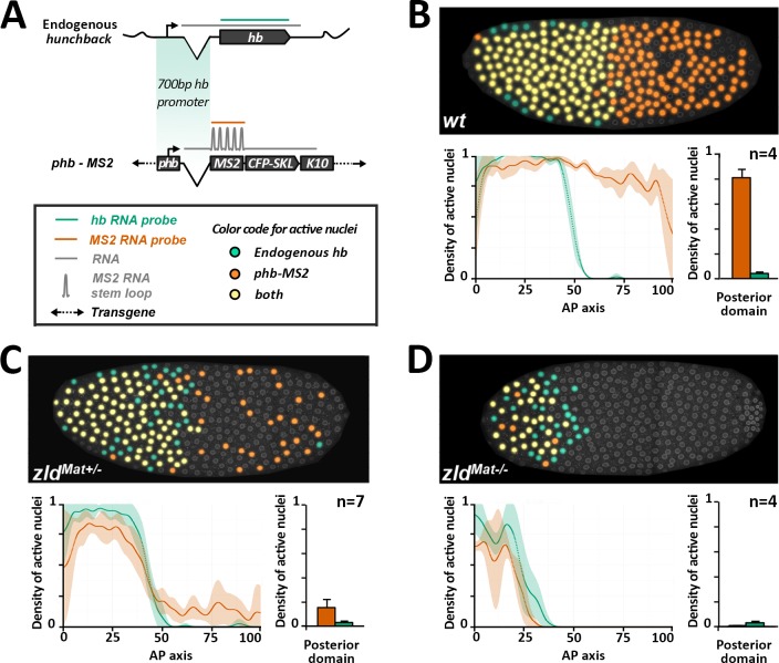
Fig 2. Posterior expression of the hb-MS2 reporters is Zelda dependent.
A) Dual RNA FISH using a hb probe (green) and an MS2 (orange) probe on wild-type embryos carrying one copy of the hb-MS2 reporter, placing the MS2-CFP-SKL-K10 cassette under the control of the 750 bp canonical promoter of hb [26]. B-D) Embryos are wild-type (B), from heterozygous mutant females for Zelda (C) or germline mutant clones for Zelda (D). Top panels: Expression map of cycle 11 embryos after segmentation of nuclei and automated processing of FISH staining. Nuclei expressing only hb are labelled in green, nuclei expressing only the hb-MS2 reporter are labelled in orange and nuclei expressing both hb and the hb-MS2 reporter are labelled in yellow. Bottom panels: On the left, density of active nuclei for either hb (green) or the hb-MS2 reporter (orange) along the AP axis with the anterior pole on the left (0) and the posterior pole on the right (100). On the right, density of active nuclei for the hb-MS2 reporter (orange) and hb (green) in the posterior domain. For each embryo, the position of the expression boundary is calculated as the position of the maximal derivative of the active nuclei density curve. Mean values were calculated for n embryos and error bars correspond to standard deviation.

