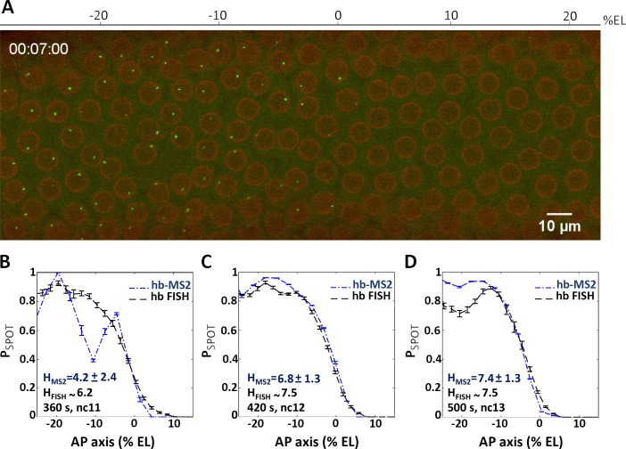
Fig 5. The hb canonical promoter expresses the MS2ΔZelda cassette in an anterior domain with a steep posterior boundary.
(A) A 2D maximum projection snapshot from Movie3 (SI) was taken at ~7 minutes after the onset of nc12 interphase. In the green channel, MCP-GFP proteins recruited by the nascent MS2-containing mRNA can be seen accumulating at the hb-MS2ΔZelda loci (bright spots). In the red channel, the mRFP-Nup proteins localized at the nuclear envelope delineate nuclei. (B-D) Probability of active hb-MS2ΔZelda loci along the AP axis (dashed blue lines with error bars), extracted from snapshots of 6 movies near the end of each nuclear cycle: 360 s after the onset of nc11 interphase (B), 420 s after the onset of nc12 interphase (C) and 500 s after the onset of nc13 interphase (D). In each panel the probability of active endogenous hb loci (black dashed lines with error bars) extracted from the FISH data from [3] is also shown. Hill coefficients (H) are indicated in blue (hb-MS2 reporter) or in black (endogenous hb from FISH data). In B, the difference between the FISH signal and the probability of active hb-MS2ΔZelda loci is due to small number of nuclei and large variability distance between them at nc11 which limit statistics.

