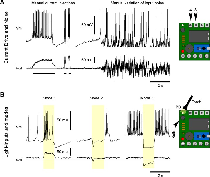
Fig 2. Manual exploration of Spikeling functions.
A. Example recording of Spikeling membrane potential (top) and current (bottom) during manual manipulations of the input current dial (4) to depolarise the neuron (left), following the addition of a noise current (dial 3, right). B. Example light responses in modes 1–3 (left to right, toggled by the button) to manual PD stimulation with a torch. The grey horizontal lines indicate Itotal = 0. PD, photoiode.

