
FIGURE 2.
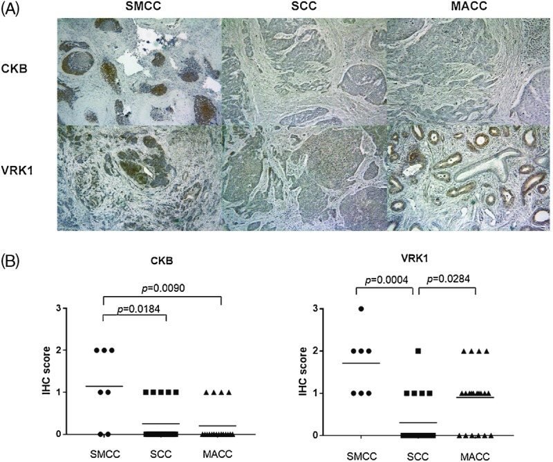
A, Representative immunohistochemical staining for CKB or VRK1 in SMCC, SCC, and MACC. Immunostained sections were counterstained with hematoxylin and photographed (magnification, ×100). The CKB was stained more intensely in SMCC than in SCC or MACC. The VRK1 was stained more intensely in SMCC and MACC than in SCC. B, IGFBP2, VRK1, and CKB immunoreactivity in SMCC, SCC, and MACC. Immunohistochemical scoring was performed according to the extent of cells staining positively.