
Figure 6.
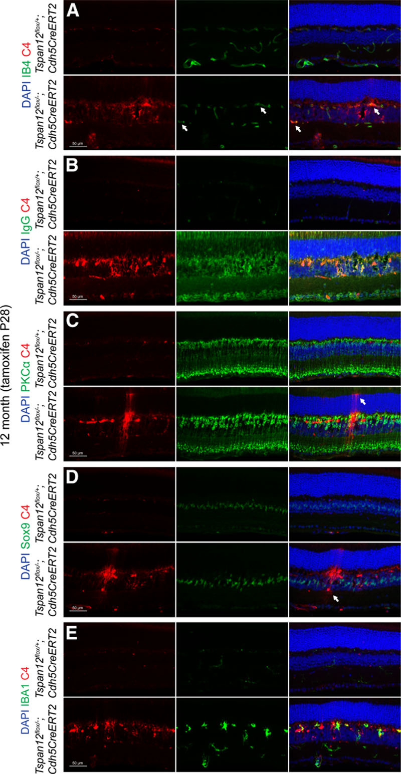
Colocalization of complement factor C4 with several retinal cell types. A, Twelve-mo-old Tspan12 ECKO retinal cross-sections stained with anti-C4 and IB4. Split and merged channels are shown. Arrows indicate blood vessels decorated with C4. B, Colocalization of extravasated IgG and C4 on a subset of retinal cells and on the borders of cystoid lesions in the mutant tissue. C, Rod-bipolar cell spacing seems increased in the mutant tissue. The section shows also 1 or 2 C4-decorated Müller glia cells, which were recognized by their extensions towards the outer limiting membrane (arrow). D, Disorganized Müller cells in the mutant tissue. Sox9 marks nuclei of Müller glia. Arrow highlights a C4-decorated cell which was recognized as amacrine cell based on cell body location and cell shape. E, Partial colocalization of C4 with microglia marker IBA-1. PKC indicates protein kinase C; and Sox9, SRY-box 9.