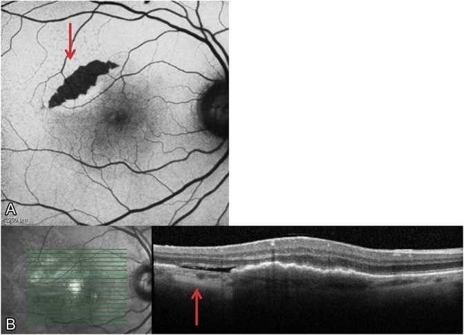
Fig. 4.

A Grade 2 RPE tear developed after several anti-VEGF injections. A. The fundus autofluorescent image illustrates the characteristic crescentic shape and hypoautofluorescent nature (red arrow) that are typical features of a tear. B. The RPE tear is also visible with SD-OCT, where areas of choroidal hypertransmission denote the absence of RPE (red arrow).
