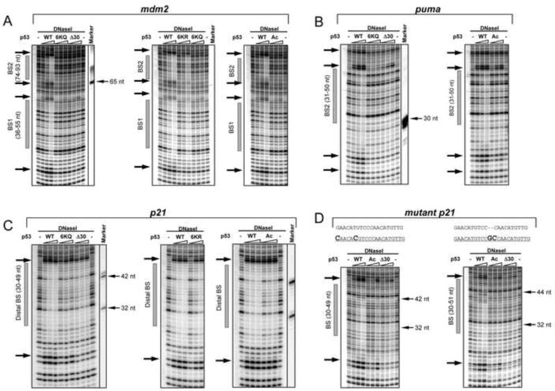
Figure 3. The p53 CTD is Required for Stable p53-DNA Complex Formation in a Sequence-Dependent Manner.
DNAseI footprint experiments with DNA fragments containing the BS from mdm2 (BS1 and BS2 weak sites, panel A), puma (intermediate site, panel B), or the p21 (strong distal BS, panel C) or mutated p21 distal BSs (panel D). The sequences of the original and two mutated p21 distal BS with changes in bold are shown on the top of gels in panel D. Each panel represents a portion of a high-resolution PhosphorImager scan of the 11 % sequencing PAAG with the positions of the corresponding BSs shown as gray bars on the left side of each scan. The arrows on the left side of each gel denote p53-specific DNaseI hypersensitive sites. Full site scanning densitometry analysis specific to each p53 variant at its maximal concentration is shown in Figure S4. Sequence-specific DNA markers were prepared and run on the gels along with the experimental reaction mixture (indicated by thin arrows on the right of side of the gel).

