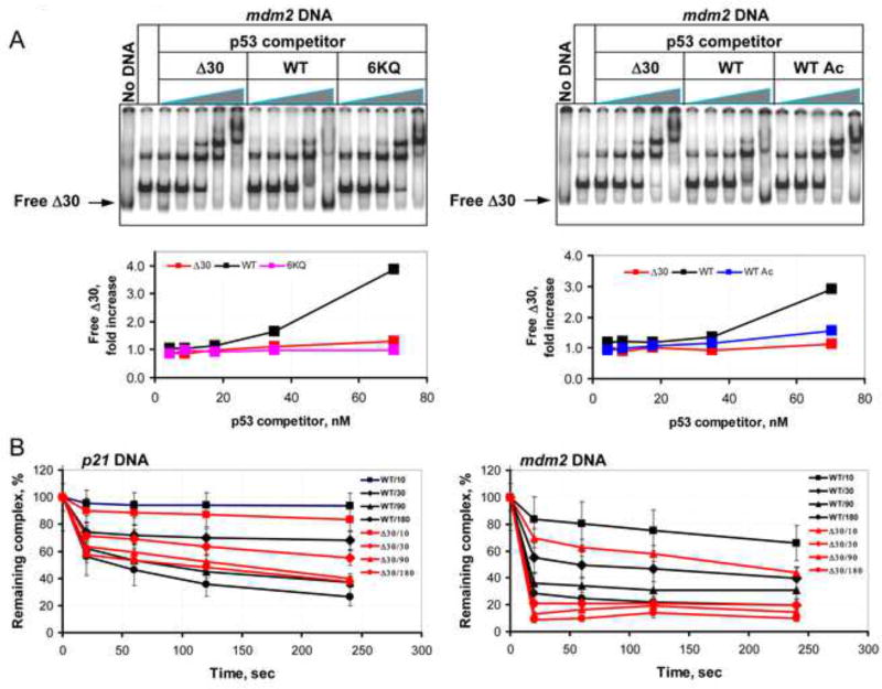
Figure 4. The p53 CTD Regulates Stability of p53-Cognate DNA Complexes.
(A). Representative gel scan of a competition binding assay that was performed with 32P-labeled Δ30 p53 protein in presence of unlabeled mdm2 BS-containing DNA and increasing amounts of unlabeled p53 WT or CTD-altered proteins as specified. The labeled Δ30 p53 protein was visualized using a PhosphorImager and relative amounts of displaced Δ30 p53 in each reaction mixture (indicated with arrow) were estimated using ImageQuant 5.2 software and presented as fold increase over the values obtained with no p53 competitor in the corresponding graphs below each scan. (B). Graphs show the quantification of the competition binding experiments performed with 32P-end-labeled p21 (left panel) or mdm2 (right panel) BS-containing DNA fragments in the presence of either unlabeled WT or Δ30 p53 protein as indicated. The experiment was performed as described in Experimental Procedures.

