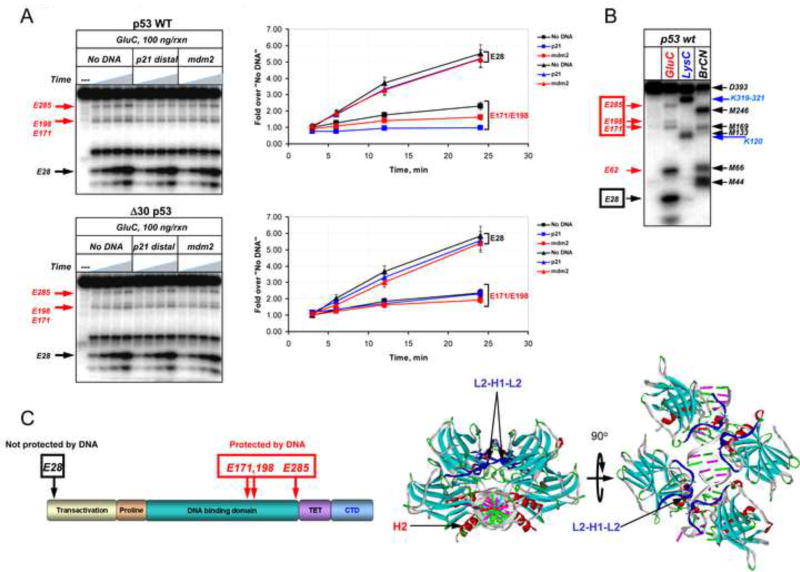
Figure 6. The p53 CTD is Important for DNA-Induced Conformational Changes within the Central Specific DNA Binding Domain.
(A). WT or Δ30 p53 proteins N-terminally labeled with 32P were incubated in the presence or absence of the indicated DNA fragments and then subjected to limited proteolysis with GluC endopeptidase for 3, 6, 12 or 24 min. The labeled cleavage products were separated by 10–20% gradient SDS TG PAGE and visualized using a Phosphorimager (left panels). The intensity of the indicated specific cleavage products of p53 was quantified using ImageQuant 5.2 (graphs on the right). (B). GluC cleavage products were identified by chemical and enzymatic mapping experiment whose details are described in Experimental Procedures using GluC, or LysC, a highly specific endopeptidase that cleaves predominantly at Lys-X; BrCN, a chemical protease that cleaves at Met-X where X is any amino acid. (C) Left: Position of the GluC cleavage sites in (A) within the full length p53 are schematically summarized in the left panel and are shown on the p53 structure (PDB 2AC0) on the right. See also Figure S6.

