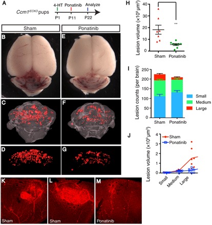
Fig. 5. Ponatinib impairs the growth of established CCM lesions in mouse model.

(A) Schematic of experimental design. Neonatal pups at P1 is induced with 4-HT and treated with ponatinib at P11. Brains and eyes were collected at P22 for micro-CT analysis and retina staining. (B to G) Micro-CT imaging of CCM lesions in brain in Ccm1iECKO with (E to G) or without (B to D) ponatinib treatment. (H to J) Quantification of micro-CT analysis shows that ponatinib treatment reduced CCM lesion volume burden by 70% (H) without changes to the total lesion number (I) in Ccm1iECKO mice compared with that of sham controls. CCM lesion distribution analysis shows decreased number of medium and large lesions in ponatinib-treated Ccm1iECKO mice, but the number of small lesions increased (I). Collective volume of small and medium lesions did not change, but the collective volume of large lesions is decreased (J). (K to M) Isolectin staining of retinal vasculature demonstrates smaller CCM lesions in the ponatinib-treated retina (M) compared with that of sham treatment (K and L). Error bars are shown as SEM, and significance was determined by Student’s t test. “**” indicates P < 0.001; n = 7 for the sham group, n = 9 for the ponatinib treatment group.
