Figure 1.

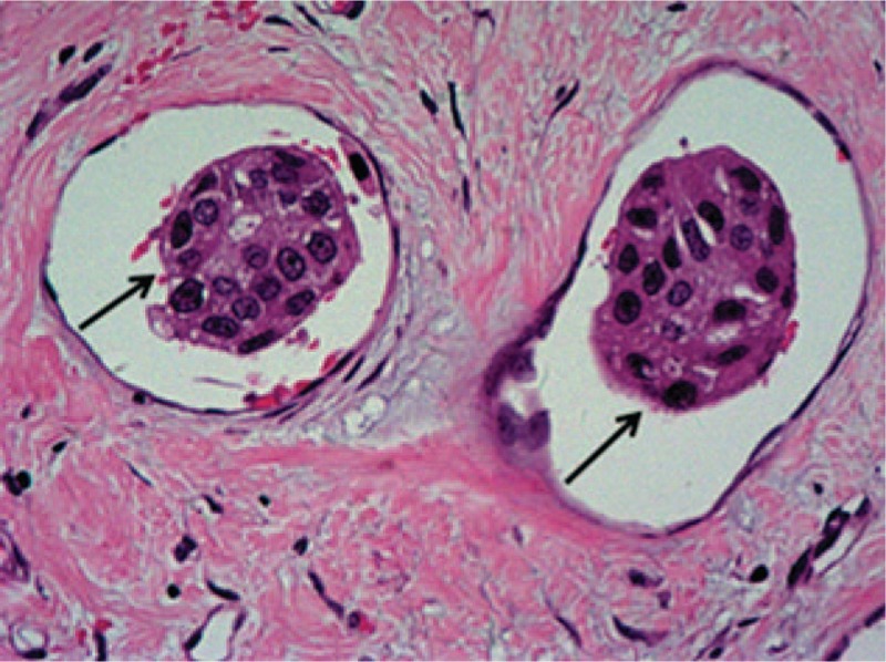
Lymphovascular invasion (LVI; single arrows) in invasive breast cancer tissue section stained with hematoxylin and eosin (H&E; magnification ×200).

Lymphovascular invasion (LVI; single arrows) in invasive breast cancer tissue section stained with hematoxylin and eosin (H&E; magnification ×200).