Figure 1.

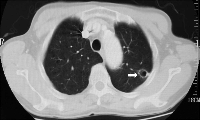
Lung-window computed tomographic image of the patient indicated an isolated thin-walled cystic lesion in left upper lobe (arrow) on November 11th, 2015.

Lung-window computed tomographic image of the patient indicated an isolated thin-walled cystic lesion in left upper lobe (arrow) on November 11th, 2015.