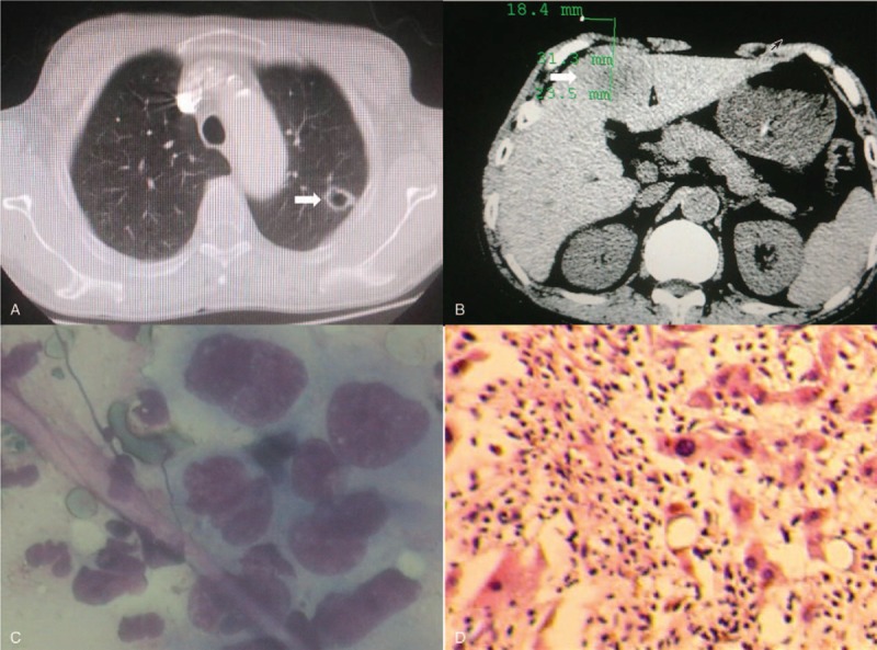
Figure 2.

Computed tomography revealed an isolated pulmonary thin-walled cystic lesion (A) and irregular liver masses (B) on June 6th, 2016. Liver biopsy displayed atypical malignant cells (C), and it was pathologically confirmed as pulmonary sarcomatoid carcinoma (D), by hematoxylin and eosin staining (×200).
