
Figure 1.
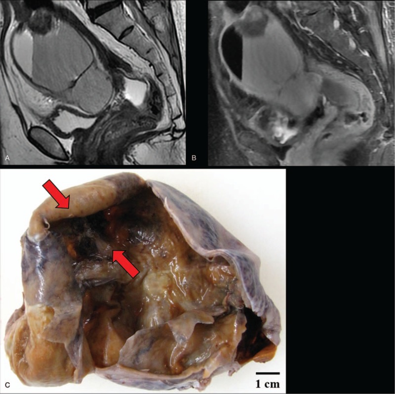
Magnetic resonance imaging and macroscopy of malignant melanoma arising in ovarian cystic teratoma: (A) T2-weighted image and (B) FAT SAT image revealed an 85 × 84 × 70-mm ovarian cystic tumor with fat. (C) The right ovarian mass had cystic appearance without solid part, but the section is darkly pigmented (red arrows) on macroscopy.