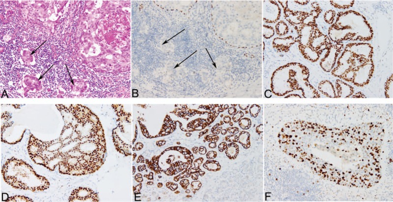
Figure 1.

Hematoxylin-eosin and immunohistochemical staining of tissues from the cases. A, Hematoxylin-eosin staining of the case with DCIS with microinvision. Two ducts are filled by ductal carcinoma in situ, while the small clusters of carcinoma cells (<1 mm) invade the stroma (arrows), 200×. B, Immunohistochemical staining for p63 highlights continuous positivity in myoepithelial cells of DCIS, while absence of myoepithelial cells around the tumor cell clusters confirms microinvasion (arrows), 200×. C–F, Immunohistochemical staining of ER (C), PR (D), HER-2 (E) and Ki-67 (F), 200×. DCIS = ductal carcinoma in situ, ER = estrogen receptor, HER-2 = human epidermal growth factor receptor 2, PR = progesterone receptor.
