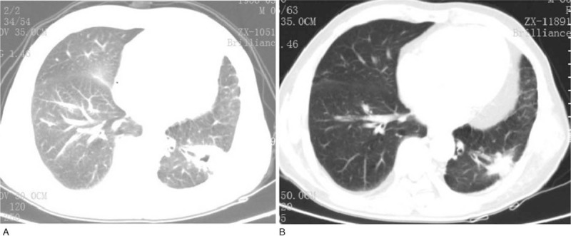
Figure 2.

Chest computed tomography scans of the patient during treatment of radiofrequency ablation. After the first time treatment of RFA, a 4.0×3.5 cm thick-walled cavities tumor is seen in the left lower lobe of the lung. Before the third time treatment of RFA, the left lung tumor decreased to 2.0×0.9 cm. RFA = radiofrequency ablation.
