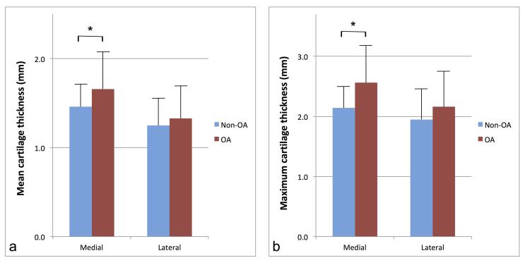
Figure 3.
Charts illustrate the comparison of the mean cartilage thickness (a) and maximum cartilage thickness (b), between non-osteoarthritis (blue) and osteoarthritis (red) groups, for both the medial and lateral condyles. Results are expressed in millimeters, and error bars correspond to standard deviations of the means. Asterisks indicate statistically significant differences.

