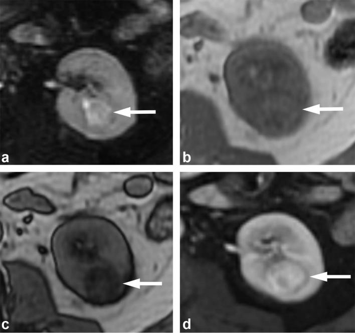
Figure 14.
A 71-year-old male with clear cell RCC. (a) T2 weighted, fat suppressed image shows the mass (arrow) has heterogeneous high T2 signal. (b) T1 weighted dual echo in-phase and (c) opposed-phase images show a left renal mass (arrows) that demonstrates signal drop in the opposed-phase image (b), consistent with the presence of microscopic fat. (d) Gadolinium-enhanced image shows avid enhancement of the mass. The combination of findings is suggestive of a clear cell renal cell carcinoma, and was proven at pathology.

