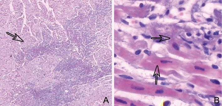
Figure 2.
H&E staining depicting infarcted myocardial lesions (arrow) and normal intersperse myocardial foci, detectable at low magnification by microscopy (a). At high magnification, myocardial cytoplasm cohesion, eosinophil level alteration, stripe disappearance, nucleus relocation, and interstitial edema of cardiomyocytes were detected (arrow) (b). H&E, hematoxylin and eosin.

