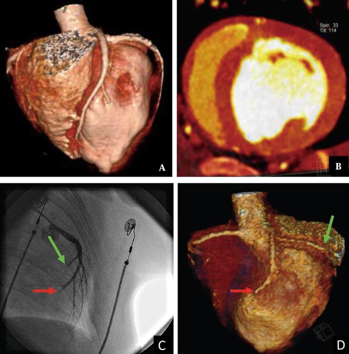
Figure 3.
Coronary CT angiography showing no occlusion in segments of the coronary artery before MI induction (a). Dual energy iodine mapping showing normal blood pool in the middle segmental region of the myocardium before MI induction (b). Invasive angiography (c) confirming obstruction in the middle of left anterior descending artery (red arrow) and left circumflex artery (green arrow). Coronary CT angiography demonstrating the obstruction (d). MI, myocardial infarction.

