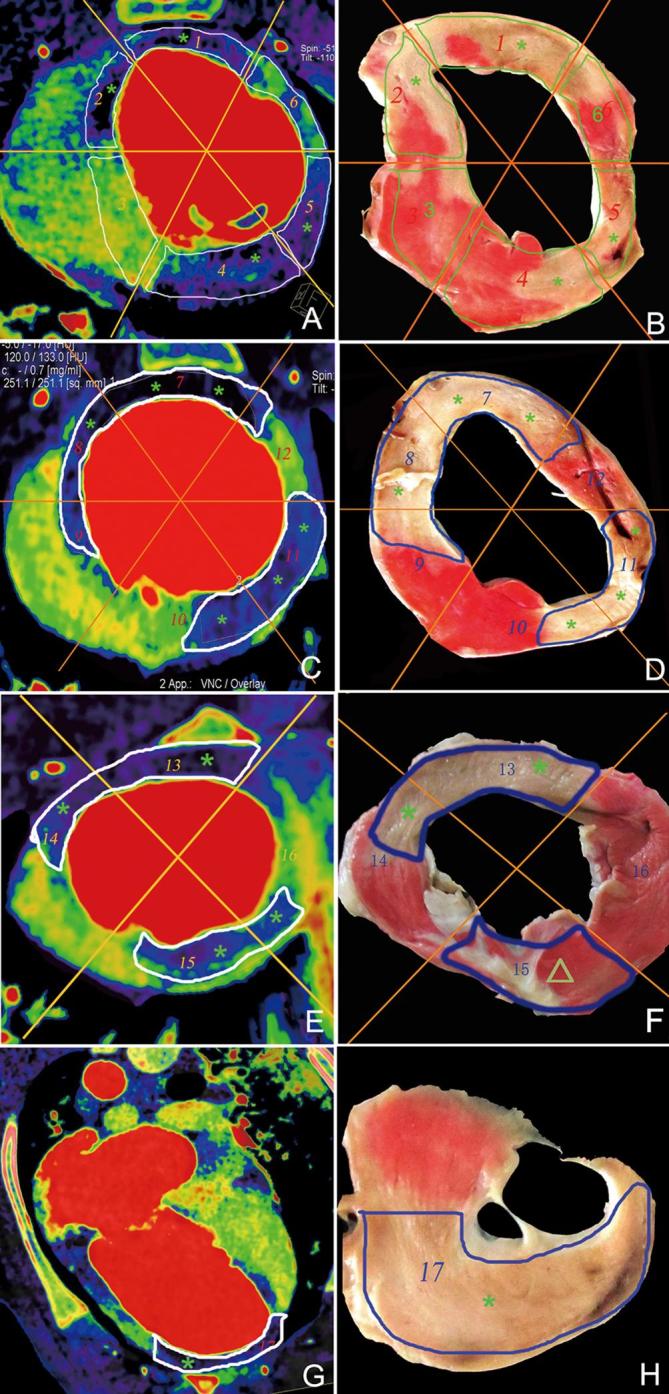
Figure 4.

Representative results of dual energy iodine mapping within the myocardial blood pool for detecting acute myocardial infarction based on pathological findings. Basal segment (a, b), middle segment (c, d), apical segment (e, f), and segments of the apex (g, h). Dual energy iodine imaging (a, c, e, and g) showing blood pool deficit in the myocardial segments 1, 2, 4, 5, 7, 8, 10, 11, 13, and 17 (*) and corresponding myocardial infarction segmental regions (*) in TTC pathology (b, d, f, and h). Myocardial segmental regions 3, 6, 9, 10, 12, 14, and 16 in dual-energy iodine imaging showed normal blood pool and corresponding normal myocardial infarction segmental regions in pathology. Dual-energy iodine imaging (e) showing blood pool deficit (*) in the myocardial segmental region 15, confirmed normal myocardial segmental region (Δ) in TTC pathology (f) (false-positive). TTC, triphenyl-tetrazolium chloride.
