Figure 1.
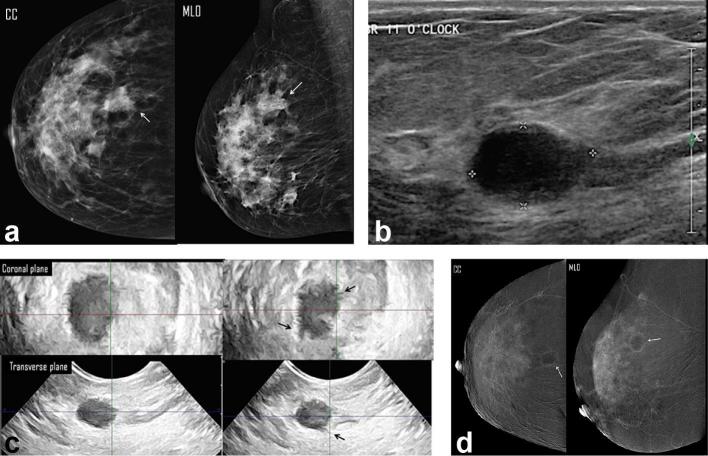
A 66-year-old female presented with right breast lump. (a) Digital mammogram MLO & CC views showed an upper outer spiculated mass associate with overlying focal dermal dimpling (white arrow) of max. dimension of 3.98 cm (overestimated length) and a nearby evident two ill-defined nodules (b) Right breast, two-dimensional ultrasound images showed three ill-defined solid masses at 12 clock; the dominant one measured 2.3 cm in max. dimension. (c) Three-dimensional breast ultrasound: coronal (left image) and transverse (right image) planes: the dominant mass was the most accessible for examination; it measured 3.5 cm, in max. dimension. No intraductal component. (d) CESM revealed an upper outer quadrant enhancing spiculated mass measured 3.5 cm in max. dimension. There were surrounding multiple enhancing smaller masses (white arrows) and foci (arrow heads). Histopathology: multifocal IDC, with max. dimension of 3.5 cm and free adherent stroma. CESM was the modality that showed accurate assessment of the multiplicity of the breast cancer. CC, craniocaudal; CESM, contrast-enhanced spectral mammography; IDC, invasive ductal carcinoma; MLO, medio lateral oblique.

