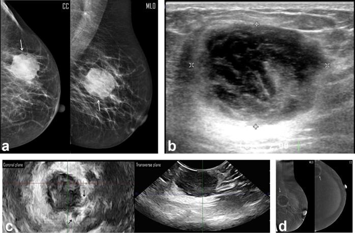
Figure 2.
A 44-year-old female presented by right breast lump. (a) Digital mammogram MLO & CC views showed heterogeneously dense breasts (ACR “c”), and an upper outer mass (white arrow) of irregular margin, not easily distinguished regarding the type of the breast density, approx. measured 3.4 cm in max. dimension. (b) Two-dimensional breast ultrasound showed complex cystic mass with posterolateral irregular thick walls and measured 1.74 cm in max. dimension. (c) Three-dimensional breast ultrasound: coronal (upper row) and transverse (lower row) planes: coronal planes revealed an indistinct margin of the mass with tiny speculations extending into the surrounding stroma. Also the transverse plane confirmed the suggestion of associated an intraductal extension (black arrows). The measured maximum dimension was 1.87 cm. (d) CESM revealed an upper outer quadrant mass with lucent center and thick non-uniform marginal contrast uptake (white arrows). No extensions into the surrounding stroma. The max. dimension measured was 2.1 cm. Note the moderate background enhancement of the breast parenchyma. Histopathology: single mass, IDC with max. dimension of 2 cm and positive surrounding stroma for cancer cell extension. CC, craniocaudal;CESM, contrast-enhanced spectral mammography; IDC, invasiveductal carcinoma; MLO, mediolateraloblique.

