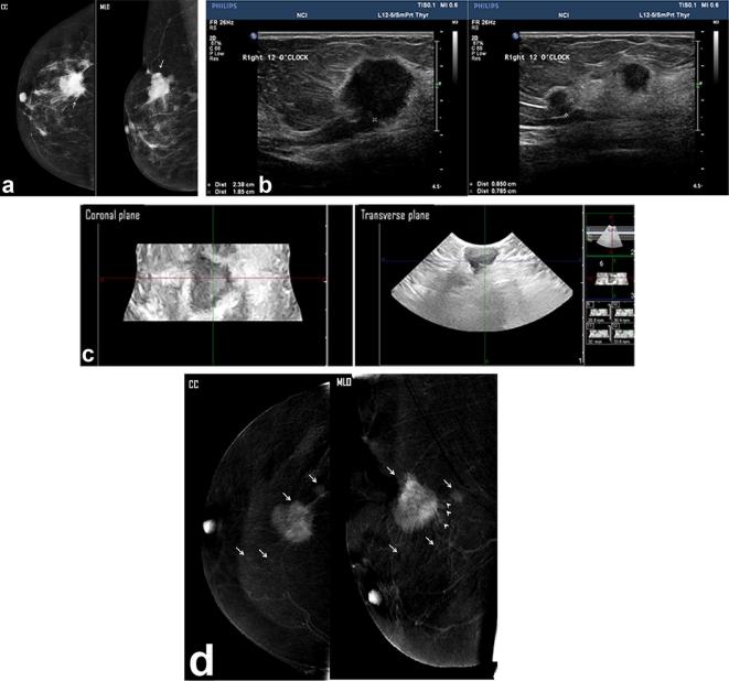
Figure 3.
A left breast lump detected in a diagnostic mammogram of a 39-year-old patient. (a) Two views digital mammogram MLO & CC showed an upper outer ill-defined mass (white arrow), mammogram overestimated the tumor size of a measured max. dimension of 5.2 cm. (b) Two- dimensional breast ultrasound showed complex cystic mass with intracystic solid components and measured in max. dimension, 3.67 cm. (c) Three- dimensional breast ultrasound: coronal (left image) and transverse (right image) planes: an intraductal component could be noted more clarified in the coronal plane (black arrows). The measured maximum dimension was 4 cm. (d) CESM revealed an upper outer quadrant mass that showed rim enhancement (white arrows). Minimal tumor extension into the surrounding stroma was noted (peri-tumoral enhancing streaks—arrows). The max. dimension measured was 4.16 cm. Histopathology: single mass, IDC with max. dimension of 4 cm and positive stromal involvement of intraductal component. Both CESM and 3D ultrasound estimated accurate mass size and positive intraductal tumor extension.

